 FIGURE 3
 FIGURE 2
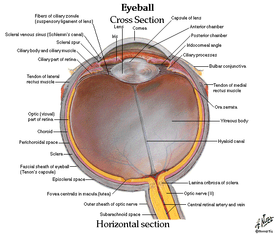
 FIGURE 1
Ladies and Gentlemen,
I bid all of you a beautiful Saturday morning and hope the day does go splendid for all of you.
For this PCL posting, I would be doing a brief summary of the anatomy of the eye but further details would be definitely be gone through during PCL this coming Monday. As usual, physiology must go hand in hand with anatomy and I believe the physiologists would be discussing together with the anatomists to bring a wholesome picture for you guys.
Gross anatomy is very dry and is just plain memory work, however, the most important thing about anatomy would be clinical relevance, so of course we would be discussing this in clinical relevance. I would just type out the pointers and everyone should go do a brief read up about the conditions. =)
For holistic sake and the non-excludability of anatomy, I would firstly be discussing about the orbit, the bones forming the orbital fossa in your skull that houses your eye. FIGURE 1
It is important to know those major bones in your skull and more importantly where glands are housed, nerves run. Like the lesser and greater wings of the sphenoid along with the floor of the maxilla and the palatine give rise to the superior orbital fissure and inferior orbital fissure. Those fissures are where some of your nerves run. The lacrimal has a fossa for the lacrimal sac and at the superior lateral aspect of your frontal bone roof has another fossa for lacrimal glands.
Clinical relevance: Blowout fracture. The medial and inferior walls of the orbit are thin and a blow may fracture the orbital walls while the margin remains intact. Orbital fractures often result in intraorbital bleeding, which exerts pressure on the eyeball, causing exopthalmos.
Lacrimal glands secrete your tears, ducts convey the tears from glands to conjunctival sac, lacrimal canaliculi each commencing at a punctum on the lacrimal papilla near the medial angle of the eye convey the lacrimal fluid from the lacrimal lake to sac, the dilated superior part of the nasolacrimal duct
Nasolacrimal duct conveys lacrimal fluid to the nasal cavity.
Production of the lacrimal fluid is by parasymp impulses from CN VII
Now we will talk about the eyelids or palpebrae (L.). When closed tit covers the eyeball anteriorly and protects it from injury or excessive light. They also help in the moisture maintenance of the eye. The eyeids are covered externally but thin skin and internally by a transparent mucous membrane called the palpebral conjunctiva. The palpebral conjunctiva is reflected onto the eyeball and continuous with the bulbar conjunctiva which is adherent to the periphery of the cornea.
The junction linking the bulbar and palpebral is known as the fornix (superior n inferior). The conjunctiva sac is the area or space bound by both palpebrae and bulbar, it allows the eyelids to move freely over the surface of the eyeball as they open and close.
The eyelashes (cilia) are in the margins of the eyelids, the large sebaceous glands associated with the eyelashes are the ciliary glands. The junctions of the superior and inferior eyelids make up the medial and lateral palpbral commissures, defining the angles of the eyes.
Do take note of the anatomy of the upper eyelids, figure 2. The tarsal glands are the ones that secrete lipids to keep the lacrimal fluid in the eye, making it moist. However when the lacrimal fluid is in too much quantity then it goes past this lipid layer and flows out as tears (when crying) cool huh!
Clinical relevance:
1. Lesion to CN III would result in ptosis.
2. Lesion to CN VII involves paralysis of the orbicularis oculi, preventing eyelids from closing fully.Loss of tonus of lower eyelid, drying up of cornea.
3.Inflammation of palpebral glands: sty and cysts
Now we will talk about the eyeball, which has 3 layers, a fibrous layer, vascular layer and a neural layer. The fibrous layer consists of the cornea and the sclera. The vascular or uvea contains iris, ciliary bodies and choroid. Neural layer consists of the retina (cones and rods).
All of these would be discussed in greater detail during the PCL.
Iris is controlled by parasymp. sphnicter pupillae and symp. dilator pupillae.
Posterior part of the eye is the fundus (hence fundoscopy)
Clinical relevance:
1.Detached retina. Seepage of fluid betwen the neual and pigmented layers of the retina, patients may complain of specks of light in front of their eye.
2.Papilledema is increase in CSF pressure slows venous return and can be seen as the swelling of the optic disc.
Remember the 4 rectus muscles, the superior and inferior oblique muscles and their movements also, what we have learnt in last semester.
Okay thats all from me for now =D Peace out people. :)))


No comments:
Post a Comment